
Double Board Certified · Functional Sinus Care
Odontogenic Sinusitis — when the cause is dental, the cure is too.
Odontogenic sinusitis is sinus inflammation caused by dental infection or dental procedure. It accounts for a meaningful share of unilateral maxillary sinusitis and is consistently under-diagnosed — because the dental and sinus evaluations are usually done by different specialists.
ABFPRS
Facial Plastic & Reconstructive Surgery
ABOto
Otolaryngology — Head & Neck Surgery
AAFPRS
Fellowship Director
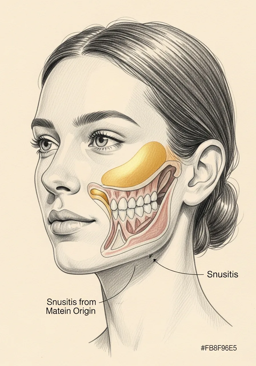
In Consultation
"Maxillary sinusitis that does not respond to standard treatment frequently has a dental cause that has been overlooked."

A Note from Dr. Mourad
"Odontogenic sinusitis is sinus inflammation caused by dental infection or dental procedure. It accounts for a meaningful share of unilateral maxillary sinusitis and is consistently under-diagnosed — because the dental and sinus evaluations are usually done by different specialists."
— Dr. Moustafa Mourad, MD
Overview
What is odontogenic sinusitis?
This page focuses on maxillary sinus disease related to a tooth or dental procedure. A dental source must be identified and treated, but dental treatment alone does not resolve every established sinus infection; some patients also require endoscopic treatment because the sinus remains obstructed, infected, scarred, or unable to clear. Compare the pattern with an acute sinus infection when symptoms begin suddenly.
Evaluation correlates one-sided maxillary findings, foul drainage, tooth or procedure history, nasal endoscopy, sinus CT, dental examination, and appropriate dental imaging. No single clue establishes the diagnosis; the sinus and dental findings should be anatomically and clinically connected.
There is no universal “dental first” or “sinus first” sequence. Dental treatment may come first when the source is clear and the sinus can recover; sinus surgery may come first or be coordinated when disease burden, blocked drainage, retained material, an oral-sinus communication, a complication, or failed prior treatment makes that appropriate.
Key takeaways
- Odontogenic sinusitis is sinus inflammation from a dental source — "odontogenic" means tooth-origin.
- Upper molar roots sit just below the maxillary sinus floor, so dental disease can spread upward.
- It is commonly one-sided and consistently under-diagnosed.
- Foul-smelling discharge or sinusitis that resists standard treatment should raise suspicion.
- Both the dental source and the sinus must be treated; sinus surgery alone is incomplete.
An Established Academic Authority
Double board certification. Fellowship director. Published author. A surgeon's surgeon.
ABFPRS
Board Certified
American Board of Facial Plastic & Reconstructive Surgery
ABOto
Board Certified
American Board of Otolaryngology — Head & Neck Surgery
AAFPRS
Fellowship Director
American Academy of Facial Plastic and Reconstructive Surgery
Textbook
Published Author
Contributions to the academic literature of facial plastic surgery
Dual board certification in both Facial Plastic & Reconstructive Surgery and Otolaryngology — Head & Neck Surgery.
Castle Connolly Top Doctor — Plastic Surgery, 202602 · Symptoms
How this condition typically presents.
Three patterns are most common. Patients often recognize themselves in one or more of these.
I
Unilateral Maxillary Symptoms
One-sided cheek pain, pressure, and congestion that does not respond to standard sinus treatment.
II
Foul Odour or Taste
A characteristic foul smell or taste — often the strongest clue to a dental cause.
III
Recent Dental History
Recent dental work, tooth extraction, or known untreated dental infection on the affected side.
03 · Anatomy
Dental & sinus anatomy.
The upper molar roots sit immediately beneath the floor of the maxillary sinus. The proximity is what makes odontogenic sinusitis possible.
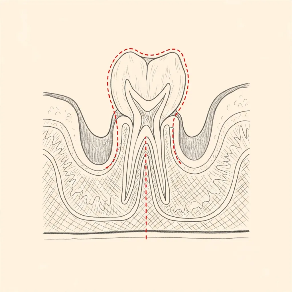
Anatomy
Root tips adjacent to the sinus floor
The roots of the upper molars frequently sit just below — sometimes within — the floor of the maxillary sinus. A periapical dental infection can extend directly into the sinus.
This anatomy is also why an upper tooth extraction can leave an oroantral communication — a small fistula between the mouth and the sinus.
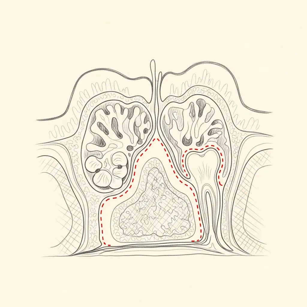
Pathology
Sinus disease from a dental source
When a dental source produces sinus disease, the imaging characteristically shows unilateral maxillary opacification with identifiable dental pathology — periapical lucency, root canal failure, or oroantral fistula.
Sinus surgery does not replace treatment of a confirmed dental source. The dental and sinus components should be assigned clearly and treated in an individualized, coordinated sequence.
Illustrative diagrams. Coordinated dental and sinus management is essential.
04 · Diagnosis
How the diagnosis is made.
Diagnosis correlates sinus CT with dental examination and appropriate dental imaging; no single clue establishes the source.
The evaluation looks for periapical disease, retained dental material, an oral-sinus communication, foreign body, or another source.
Nasal endoscopy assesses corresponding drainage and the sinus pathway so the dental and sinus findings can be connected.
05 · Treatment Options
Treatments matched to the diagnosis.
Treatment is individual. The right answer ranges from optimized medical therapy to a focused procedure to definitive surgery.
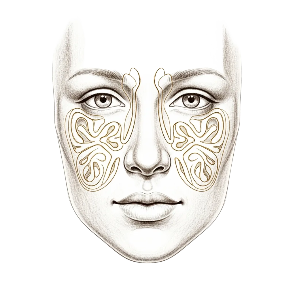
Dental Source Management
Treatment of the underlying dental cause — extraction, root canal, or repair of an oroantral fistula.
Learn More
Endoscopic Sinus Surgery
Drainage and clearance of the affected sinus, typically performed after or with dental management.
Learn More

Medical Therapy
Targeted antibiotics, saline irrigation, and supportive care as part of combined management.
Learn More
01 · Why Dr. Mourad
Diagnosis first, treatment second.
Dr. Mourad coordinates evaluation with dental specialists when odontogenic sinusitis is suspected — both the sinus and the source must be addressed.
CT imaging is essential to identify dental pathology and to plan combined treatment.
Sinus surgery alone — without addressing the dental source — produces incomplete and short-lived improvement.
When to Seek Care
When to seek care promptly.
Severe facial pain, swelling, or fever — evaluate within days.
Significant facial swelling or redness around the eye — evaluate immediately.
Numbness of the cheek or upper teeth — evaluate promptly.
Persistent oroantral fistula after recent dental extraction — evaluate within days.
Outlook
What to expect.
When the diagnosis is correct and the right treatment is applied, the outlook is generally good. Most patients describe meaningful improvement in symptoms and day-to-day function.
When symptoms persist despite treatment, the workup is re-opened. Persistent symptoms with no answer almost always mean the diagnosis is incomplete.

Living Well
Day-to-day measures that help.
Daily saline irrigation, control of indoor allergens, and good sleep hygiene meaningfully reduce day-to-day symptoms for many patients.
Medical therapy, when prescribed, works best when used consistently rather than as needed — this is one of the most common reasons treatment seems to fail.
Frequently Asked
Patient questions, honestly answered.
Yes. An infected upper tooth, failed root canal, extraction, implant, or communication between the mouth and sinus can infect the adjacent maxillary sinus. One-sided symptoms and a bad smell or taste are clues.
Yes. The tooth may be dead, previously treated, or only mildly symptomatic while the sinus causes congestion, pressure, foul drainage, or a bad taste.
Continue Reading
Related sinus care
The Most Important Step
Get an expert evaluation.
A careful evaluation by a double board-certified physician is the right first step. The conversation is unhurried, the diagnosis is honest, and treatment is matched to what you actually have.