Double Board Certified · Specialized Revision Practice
Revision Rhinoplasty NYC — Correct, Rebuild, Restore
Dr. Moustafa Mourad performs revision rhinoplasty in NYC for patients whose nose looks, breathes, or feels different than intended after prior surgery. Revision planning begins by identifying what was removed, what weakened, what scarred, what remains available, and whether the internal airway was affected. The operation may involve scar release, septal or valve reconstruction, cartilage grafting, implant or graft management, and conservative reshaping to restore support before further refinement.
ABFPRS
Facial Plastic & Reconstructive Surgery
ABOto
Otolaryngology — Head & Neck Surgery
AAFPRS
Fellowship Director

In Consultation
"The hardest cases are the ones I am sent."
Before & After
Revision Rhinoplasty Before and After Photos
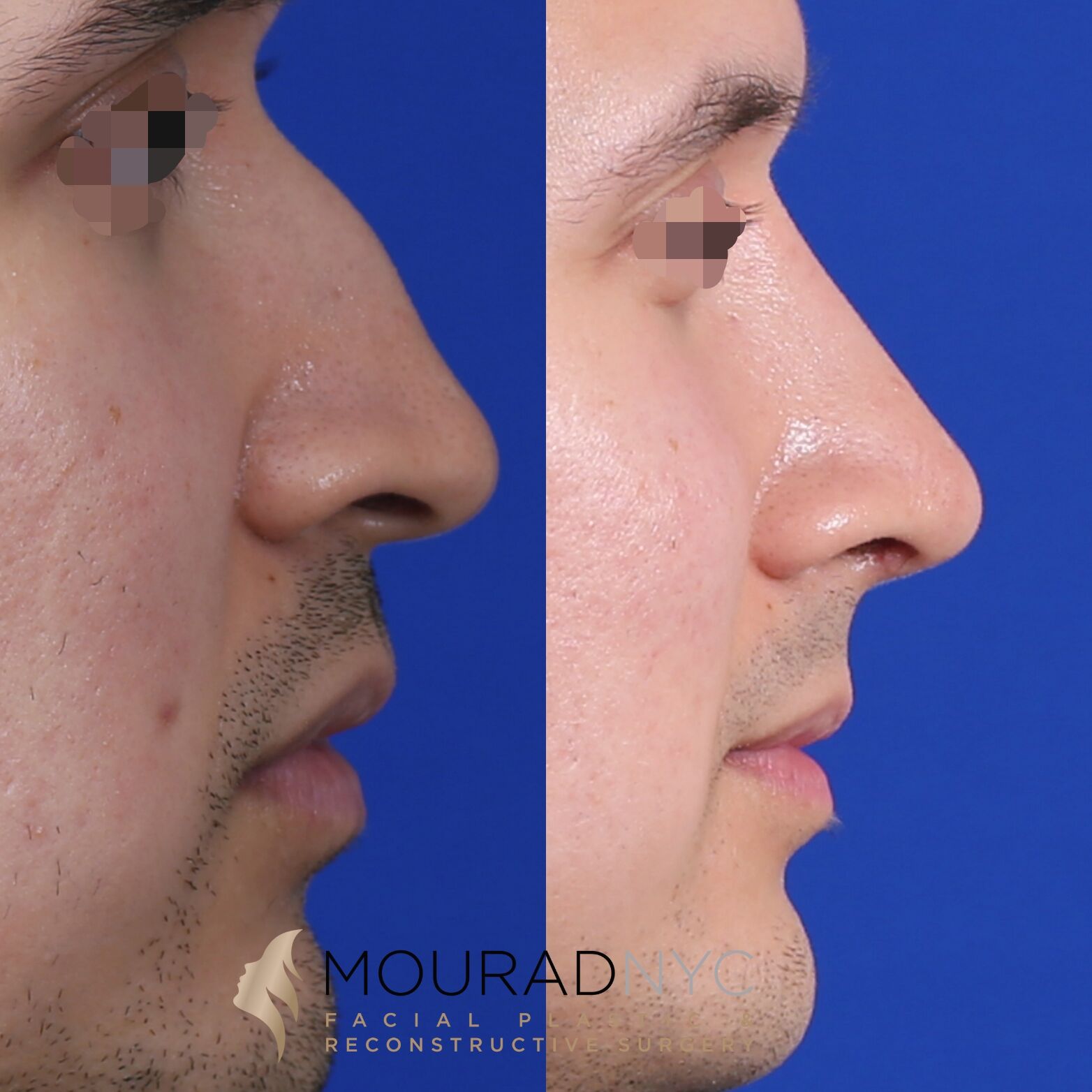
Revision Rhinoplasty · 30s · Male
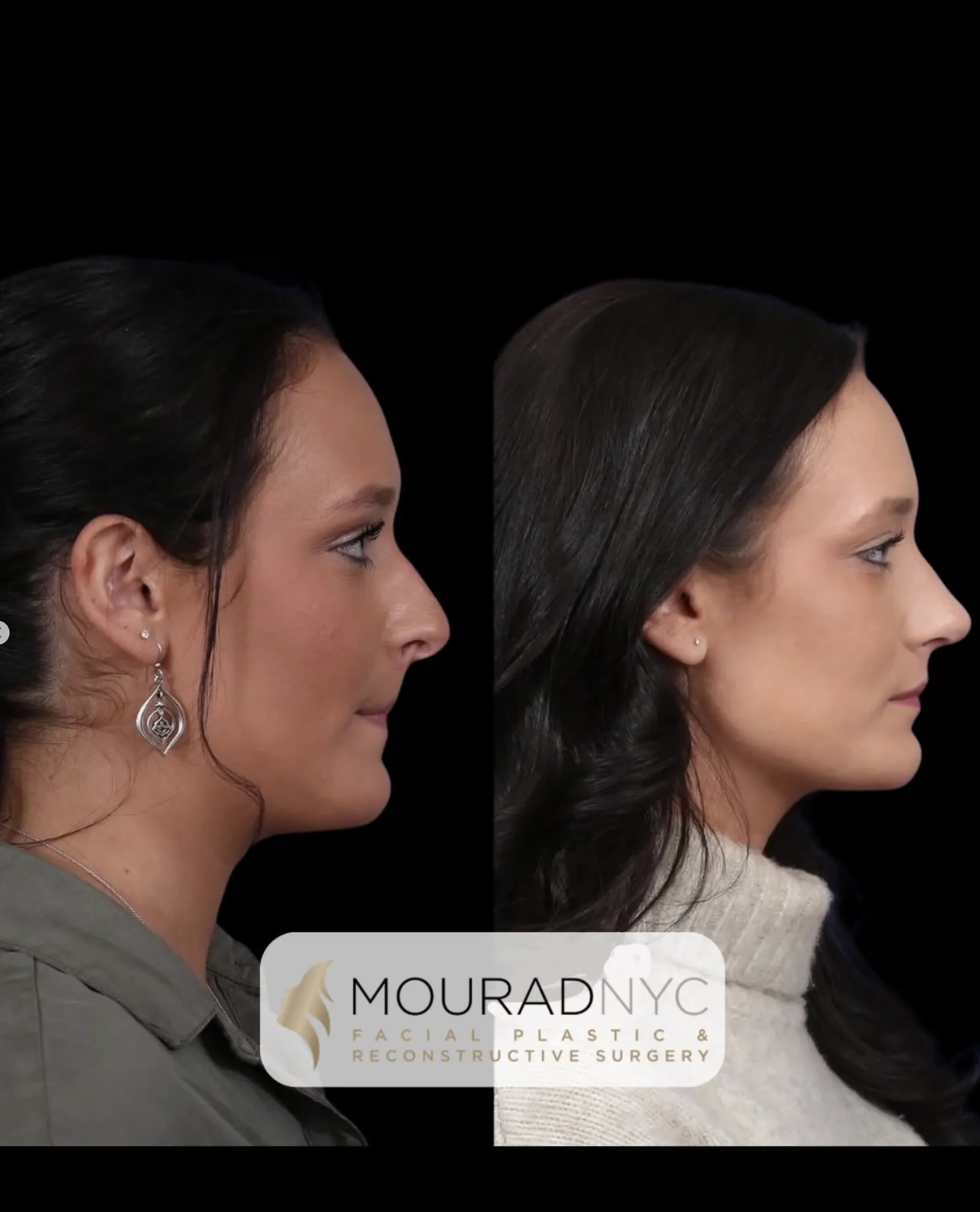
Revision Rhinoplasty · 20s · Female
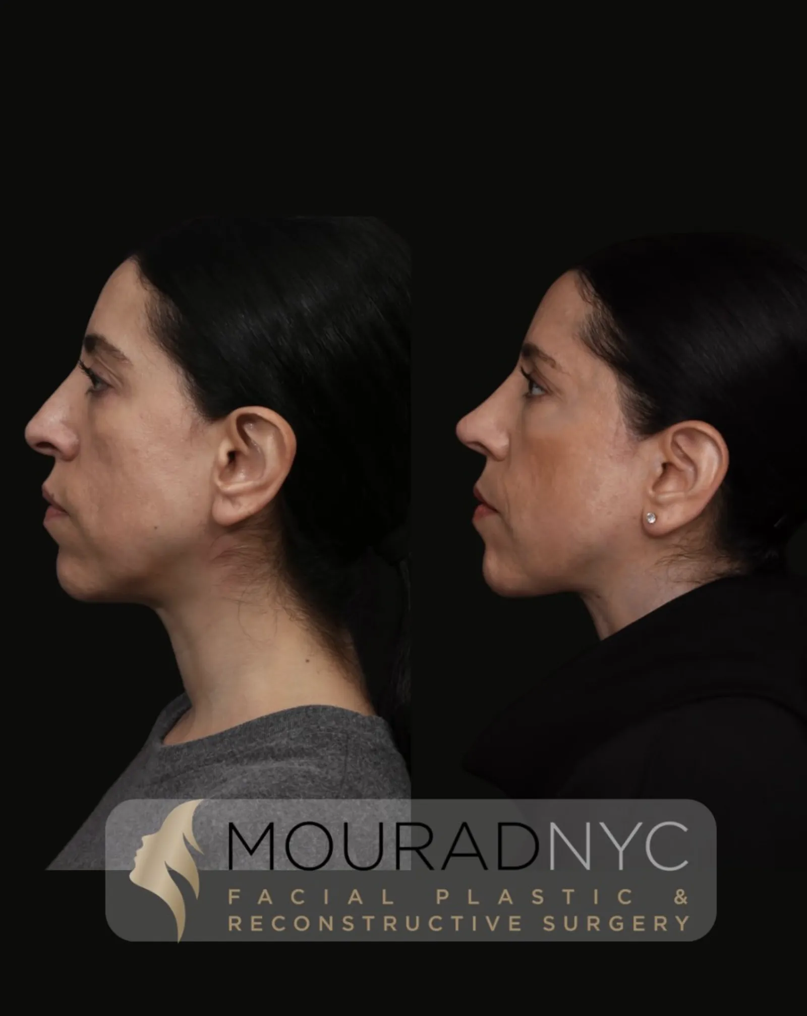
Revision Rhinoplasty · 30s · Female
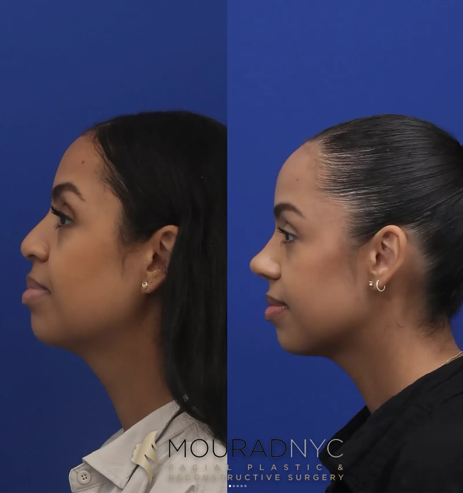
Revision Rhinoplasty · 30s · Female
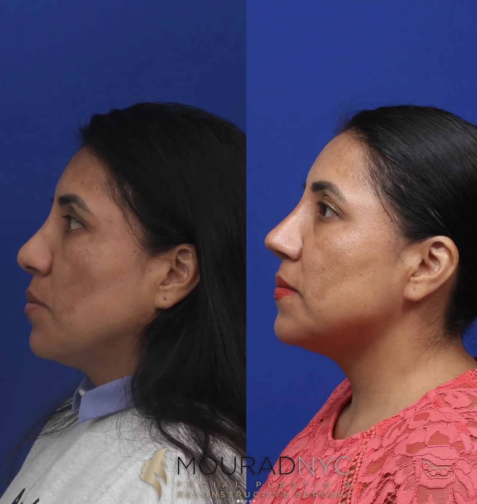
Revision Rhinoplasty · 40s · Female
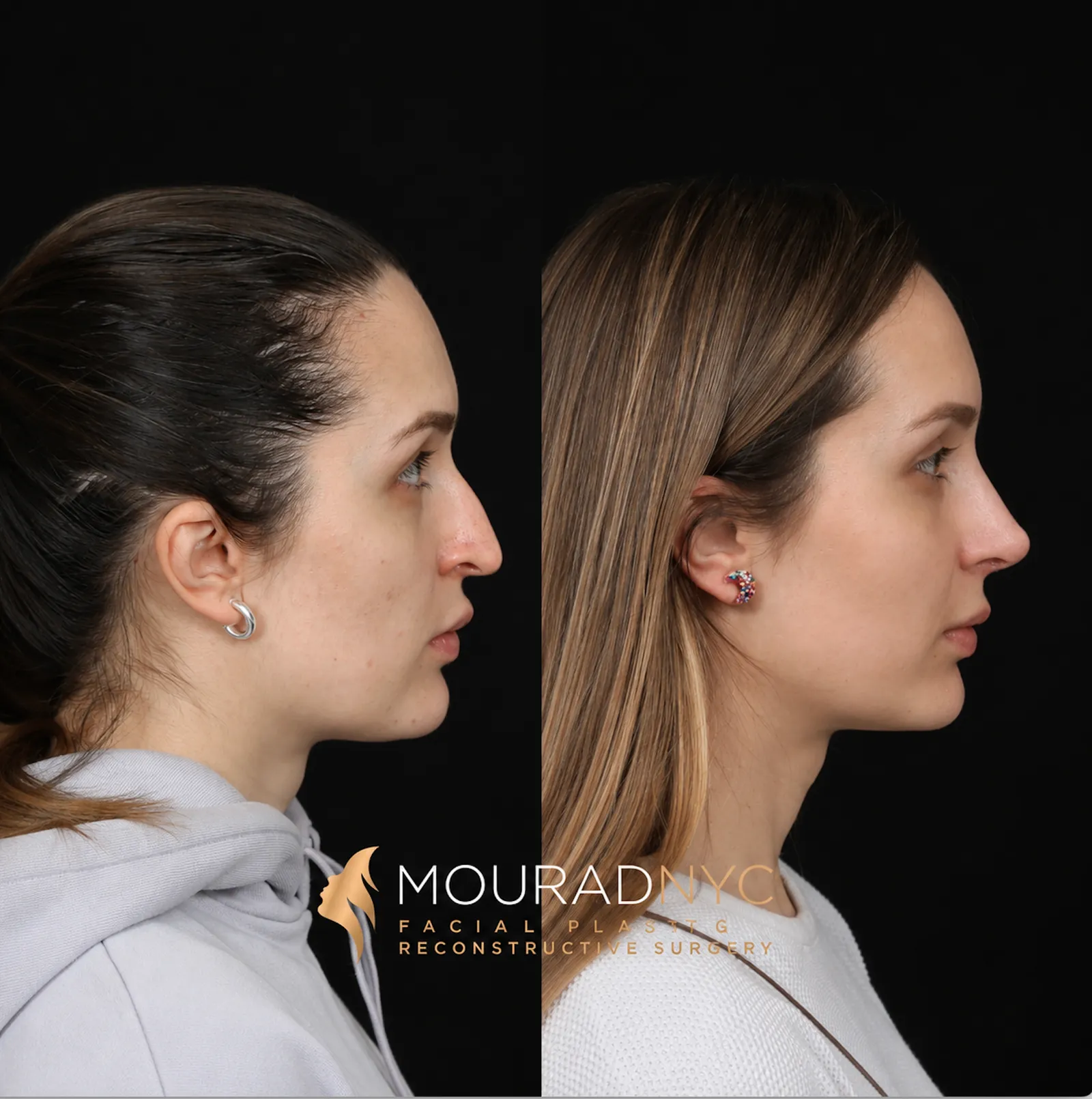
Revision Rhinoplasty · 30s · Female
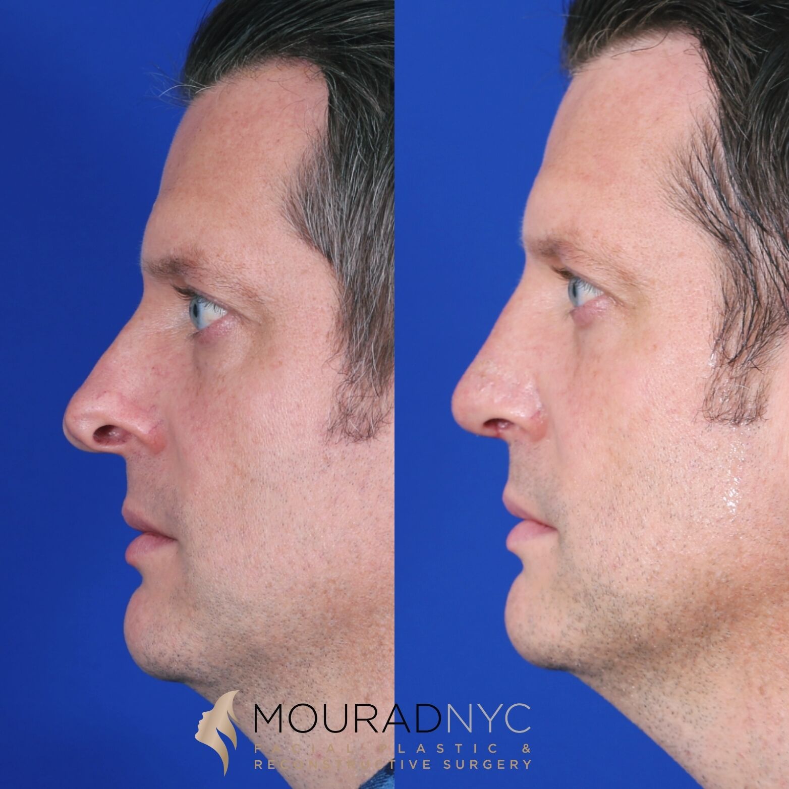
Revision Rhinoplasty + Sinus Surgery · 30s · Male
Pre and post-operative comparison · Photographed in standardized studio conditions · Written consent on file · Individual results vary

A Note from Dr. Mourad
"Revision rhinoplasty is a different operation from primary rhinoplasty. The scar tissue is dense, the cartilage is depleted, and the airway has often been compromised. What this work demands is restraint, structural thinking, and a willingness to take cases that others decline."
— Dr. Moustafa Mourad, MD
Medically reviewed by Moustafa Mourad, MD, FACS — dual board-certified in Facial Plastic & Reconstructive Surgery and Otolaryngology–Head & Neck Surgery; Clinical Assistant Professor of Otolaryngology at New York Medical College; AAFPRS Fellowship Director. The medical review identifies the anatomy, evidence, limits, and questions patients should understand before choosing a procedure.
Last reviewed: June 2026
Make sure you're in the right place
Is this the right page for you?
- You have already had one or more nasal surgeries and remain unhappy with the breathing, the appearance, or both.
- You have scar tissue, weakened or depleted cartilage, or a nose that looks or functions differently than planned after prior surgery.
- You were told elsewhere that your case is too complex or inoperable.
You may be looking for
- If you have never had nasal surgery, primary rhinoplasty is the right starting point.
- If your only concern is breathing through a deviated septum, see septoplasty.
- If your nose was recently broken, see broken nose repair.
Key takeaways
- Revision rhinoplasty corrects a nose after prior surgery, where scar tissue and depleted cartilage add complexity.
- Cartilage grafts — from the septum, ear, or rib — rebuild lost support before refinement is possible.
- Timing is individualized: many elective revisions wait for swelling and scar to mature, while a defined functional or infectious problem may warrant earlier care.
- Breathing and appearance are addressed together, since the structural and aesthetic fixes overlap.
- Not every concern can be fully corrected; realistic goals depend on anatomy, scarring, and healing.
Overview
What is revision rhinoplasty?
This page focuses on surgery after a prior rhinoplasty. Patients researching a first operation should begin with Primary Rhinoplasty or the flagship Rhinoplasty NYC overview.
Revision rhinoplasty is rhinoplasty performed on a nose that has already had previous nasal surgery. Sometimes called secondary rhinoplasty or corrective nose surgery, it is an inherently more complex operation than primary rhinoplasty because the anatomy has been altered, the cartilage stock may be depleted, scar tissue distorts normal tissue planes, and the underlying structural support may be weakened by prior reduction. Access may involve open vs. closed rhinoplasty, chosen for the anatomy and reconstruction required.
Patients pursue revision for many reasons — residual cosmetic concerns, asymmetry, an over-rotated or pinched tip, a saddle deformity, a pollybeak fullness, or new breathing problems that emerged after the first operation. Functional revision and cosmetic revision are frequently performed together because the structural fix and the aesthetic fix are the same operation.
A revision plan may require remaining septal cartilage, ear cartilage, rib cartilage, fascia, or another reconstructive material to restore support before refinement. No source is automatic. Graft choice and timing depend on the defect, available tissue, skin and scar condition, airway findings, donor-site considerations, and whether the prior operation has healed enough for safe re-entry. Patients should also understand the considerations in how to choose a rhinoplasty surgeon.
What makes revision rhinoplasty different from a first operation?
Revision rhinoplasty is surgery on a nose that has already been operated on. Scar tissue, altered anatomy, and reduced cartilage make it more complex than a primary procedure, and rebuilding structural support with cartilage grafts is often part of the plan.
A careful evaluation of what the previous surgery changed — and what tissue remains to work with — comes before any plan, which is why these cases are approached more conservatively than a first rhinoplasty.
When is rib cartilage needed in revision rhinoplasty?
Rib cartilage may be considered when the reconstruction requires more or stronger material than the remaining septum or ear can provide. It can supply structural grafts for the bridge, tip, septum, or sidewalls, but a prior operation alone does not make rib cartilage necessary.
Whether rib, ear, or remaining septal cartilage is the right source depends on how much structure needs to be rebuilt, which is assessed at consultation and sometimes with imaging.
See a rib-graft caseWhat problems can revision rhinoplasty address?
Revision rhinoplasty can address structural collapse, breathing obstruction, asymmetry, over-resection, a pinched or upturned tip, and irregularities along the bridge from a prior operation. Many revisions combine airway repair with refinement of the external shape.
The realistic scope of a revision depends on the quality of the remaining tissue — some concerns can be improved substantially while others are managed more conservatively.
How long should I wait before revision rhinoplasty?
Many elective revisions are delayed until swelling has settled and scar has matured, commonly around a year but not according to one universal calendar. Earlier intervention may occasionally be appropriate for infection, exposed material, severe obstruction, displacement, or another defined problem.
The right timing is individualized — an earlier consultation can still be useful to document the anatomy and plan ahead, even if surgery itself waits.

BEST of MANHATTAN
2026
Modern Luxury Best of Manhattan 2026
Rhinoplasty recognition, applied carefully to complex revision planning
Dr. Moustafa Mourad was named "Best Rhinoplasty" in Modern Luxury's Best of Manhattan 2026 medical category. For revision patients, the relevance is not a slogan — it is the same rhinoplasty judgment applied to scar tissue, altered anatomy, airway compromise, and structural rebuilding.
Third-party recognition is not a guarantee of outcome. Every rhinoplasty plan is individualized.
Diagnosis Before Technique
Revision diagnosis comes before revision technique
A revision consultation should identify the cause of the problem before discussing an operation. Similar-looking deformities may have very different structural causes: supratip fullness can reflect scar, residual cartilage, or weak tip support, while a narrow middle vault can reflect resection, displacement, scar, or inadequate reconstruction. The safest correction depends on the tissue that remains rather than the appearance alone.
Diagnosis combines the patient's timeline with external examination, airway assessment, standardized photographs, and prior records when available. Endoscopy or selective imaging may be useful for a suspected septal perforation, scar narrowing, implant issue, sinus finding, or another internal concern, but neither automatically replaces a hands-on examination.
- —What did the prior operation change? Review visible contour, septal support, tip position, bridge height, nostril shape, and airway function.
- —Is the problem tissue loss, scar, displacement, or incomplete correction? Each mechanism requires a different plan and may impose different limits.
- —What cartilage remains? Septal reserve, prior grafts, ear or rib options, fascia, and any implanted material affect feasibility.
- —How healthy is the skin and soft-tissue envelope? Thin, scarred, inflamed, or poorly vascularized tissue limits how aggressively the framework can be changed.
- —Is breathing impaired? Residual septal deviation, internal or external valve collapse, turbinate disease, and scar narrowing should be separated from purely cosmetic concerns.
- —Has the nose healed enough for another operation? Timing depends on swelling, scar maturity, inflammation, infection, functional urgency, and the reason for revision.
What It Corrects
What revision rhinoplasty may need to correct
Revision rhinoplasty addresses a defined set of problems left behind by a prior operation. The right plan depends on which of these groups applies and on what the tissues will now tolerate.
- —Persistent shape concern — the original concern remains or was incompletely corrected.
- —Over-resection — excessive removal may create a pinched tip, inverted-V deformity, saddle nose, alar retraction, or valve narrowing.
- —Under-resection or scar contour — residual hump, pollybeak, asymmetry, or scar-related fullness may remain.
- —Support loss — the tip, middle vault, nostril rim, or sidewall may lack stable cartilage support.
- —Breathing problems — the septum, valves, turbinates, scar, or prior structural changes may contribute.
- —Graft or implant problem — prior grafts or synthetic materials may be visible, displaced, infected, or poorly integrated.
Functional Evaluation
Breathing problems after prior rhinoplasty
Nasal obstruction after prior surgery can arise from more than one structure. Residual septal deviation, internal or external nasal-valve collapse, turbinate enlargement, scar narrowing, loss of middle-vault support, over-resection, or a displaced graft may produce similar symptoms.
The revision examination separates these causes before another operation is planned. A revision may include septal correction, valve reconstruction, scar release, turbinate treatment, or restoration of structural support, but none of these steps is added automatically. The operation should treat the documented source of obstruction and account for how functional reconstruction will affect the external contour.
Patients whose concern is breathing alone may be better served by a focused functional page or procedure. Patients with both contour and airway problems require one coordinated revision plan.
How Dr. Mourad Plans
How Dr. Mourad plans a revision rhinoplasty
Revision planning begins with the patient's story. What was the original goal? What changed after surgery? Did breathing worsen? Did the shape change as swelling resolved? Was there trauma after surgery? Were implants or grafts used? Was there infection, prolonged swelling, filler, or steroid injection?
The next step is photographic review. Old photos can show the original anatomy and help separate preexisting asymmetry from surgery-related changes. Operative reports are valuable because they may reveal whether septal cartilage was removed, whether spreader grafts were placed, whether osteotomies were performed, and whether alloplastic materials or cadaveric cartilage were used.
The examination evaluates both appearance and airway — the bridge, middle vault, tip, nostril rims, alar base, columella, septum, turbinates, internal and external valves, skin thickness, scar density, and cartilage support. Nasal endoscopy may be used to assess the septum, valves, turbinates, perforation, or synechiae.
The surgeon then determines the cartilage inventory. If septal cartilage remains, it may be useful; if not, ear or rib cartilage may be needed. Major support loss, saddle deformity, valve collapse, or multiple prior surgeries often require stronger cartilage than the septum or ear can provide.
The final plan prioritizes safety. Sometimes the patient should wait longer before revision; sometimes staging is safer than attempting every correction at once. In all cases revision is less predictable than first-time rhinoplasty, and improvement — not perfection — is the goal. For context on the operation as a whole, see the rhinoplasty overview.
Implant & Graft Complications
Revision after an implant, cadaveric graft, or prior graft failure
Some revision patients have prior grafts or implants that no longer behave as intended. These may include silicone, Gore-Tex, Medpor, cadaveric rib cartilage, irradiated cartilage, ear cartilage, septal cartilage, or rib cartilage from a previous operation. The problem may be infection, exposure, extrusion, resorption, warping, displacement, contour visibility, skin thinning, or collapse after support weakens.
A prior implant does not automatically mean it must be removed, and a prior cadaveric graft does not automatically mean it failed. The decision depends on symptoms, skin quality, infection risk, stability, contour, breathing, and whether the material is contributing to the problem.
Silicone, Gore-Tex, and Medpor may create special concerns because they are foreign materials. If they become infected, exposed, mobile, or visible through the skin, removal and reconstruction may be needed. In some patients the surrounding soft tissue may be thin or scarred, which can make revision more complex.
Cadaveric rib cartilage can be useful in selected patients, but if it resorbs, warps, shifts, or fails to provide durable support, the nose may need reconstruction with autologous cartilage. Autologous rib cartilage may be preferred when strong structural support is required, especially in major saddle nose repair, severe middle-vault collapse, or revision after multiple prior operations.
For major structural reconstruction and complex revision rhinoplasty, autologous rib cartilage is generally favored when substantial strength, volume, and long-term support are required. It provides abundant graft material with predictable structural characteristics but requires a separate chest donor site. Cadaveric rib can be appropriate in selected patients because it avoids a chest incision and donor-site morbidity; the choice is individualized based on the reconstructive demands and patient preferences.
A sound revision plan begins with identifying what is currently in the nose. Operative reports, prior imaging, old photos, and a careful exam all help. Patients should bring any available operative records to consultation. For implant-specific planning, read about revision rhinoplasty after implants, Gore-Tex, or silicone.
Timing
When revision should be delayed
Many elective revisions are delayed until the prior operation has had enough time to settle and the scar has matured, commonly around a year but not according to one universal calendar. Operating while inflammation or rapid change continues can obscure the true problem, make dissection more difficult, and lead to treatment of a contour that would otherwise improve.
Earlier evaluation is useful even when surgery is deferred. Earlier intervention may occasionally be appropriate for infection, exposed material, severe obstruction, displacement, trauma, or another defined problem. Timing is determined by examination, tissue condition, the pace of healing, and the reason for revision.
Patients should be cautious about chasing early swelling. Irregularities seen at three or six months may improve. Conversely, true structural collapse or valve compromise may become more obvious as swelling resolves. Timing must be chosen based on anatomy, healing, and risk.
Records & Timing
Records to bring and how timing is decided
Bring operative reports, prior photographs, implant information, and pathology or imaging when available. These records help identify what was removed, what was added, and what remains available for reconstruction.
Revision timing depends on the reason for surgery, tissue condition, inflammation, and scar maturation. Many elective revisions are delayed until the nose has had adequate time to heal, but urgent functional or infectious problems require individualized evaluation. There is no single, rigid waiting period that applies to every patient.
When Restraint Is Safer
When another operation is not the right next step
Not every persistent irregularity after rhinoplasty should be revised. Early swelling, scar firmness, temporary asymmetry, and changing tip position may continue to evolve for many months. Operating before the anatomy stabilizes can make diagnosis and correction less predictable.
Dr. Mourad may recommend observation, medical treatment, steroid treatment when appropriate, filler dissolution when relevant, additional records, imaging, psychological support, or no further surgery. A revision is recommended only when there is a defined problem, a realistic opportunity for improvement, adequate tissue or graft options, and an acceptable balance between benefit and risk.
Some noses cannot be restored to an untouched state. The goal is meaningful improvement within the limits created by prior surgery, scar tissue, skin quality, airway damage, and remaining cartilage.
- —Which concerns can realistically improve, and which may remain?
- —Is structural or functional repair more important than additional reduction?
- —What donor tissue or camouflage may be needed, and what tradeoffs follow?
- —Do scar, skin, inflammation, or vascular limitations argue for restraint or staging?
- —Does the likely benefit justify the added risk and recovery of another operation?
What It Can Address
Revision Rhinoplasty Problem Map
Revision rhinoplasty addresses a defined set of problems left behind by prior nasal surgery. The right plan depends on what was done before and what the tissues will now tolerate. Where a dedicated page exists, each card links to a more detailed explanation.
Pinched tip
A pinched tip may occur when too much lower lateral cartilage has been removed, weakened, or scarred. The nostril rims can look narrow, the tip may appear sharp or collapsed, and the external nasal valve may not support airflow. Revision often requires rebuilding support with cartilage grafts rather than simply reshaping the surface.
Pinched nose after rhinoplasty →Pollybeak deformity
Pollybeak deformity describes fullness in the supratip region, where the area above the tip looks convex or beak-like. It may result from residual cartilage, scar tissue, weak tip support, or disproportion between the bridge and tip. Treatment depends on whether the fullness is scar, cartilage, skin thickness, or structural underprojection.
Pollybeak deformity →Saddle nose deformity
A saddle nose occurs when the bridge loses support and becomes depressed. It may follow septal over-resection, trauma, infection, autoimmune disease, septal perforation, or prior surgery. Revision usually requires structural rebuilding, often with rib cartilage when support loss is significant.
Saddle nose deformity →Inverted-V deformity
An inverted-V deformity can appear when the middle vault collapses after hump reduction or inadequate support of the upper lateral cartilages. Patients may see visible shadowing along the upper nose and may also have internal nasal valve obstruction. Spreader grafts or other middle-vault reconstruction may be needed.
Inverted-V deformity →Alar retraction
Alar retraction occurs when the nostril rim sits too high or pulls upward, creating excessive nostril show or asymmetry. It may result from cartilage over-resection, scar contraction, prior surgery, or weak lower lateral cartilage support. Correction may require alar rim grafts, composite grafts, or structural reconstruction.
Alar retraction →Over-rotated tip
An over-rotated tip may make the nose look too upturned or expose too much nostril. In revision this can be difficult because the skin and scar tissue may resist repositioning. Correction may require tip support, septal extension grafting, or other structural maneuvers.
Residual hump
A residual hump may remain after primary rhinoplasty or become visible after swelling resolves. The cause may be residual bone, cartilage, callus, or imbalance between the radix, dorsum, and tip. Revision should be conservative and precise, because additional dorsal work can affect nasal stability and valve function.
Crooked nose after surgery
A nose may remain crooked or become crooked after surgery because of bone memory, cartilage memory, septal deviation, asymmetric scarring, or incomplete correction of the original deformity. Revision requires identifying whether the deviation is bony, cartilaginous, septal, soft tissue, or facial asymmetry.
Nasal valve collapse
Nasal valve collapse after rhinoplasty may occur when the middle vault or lower lateral cartilages lose support. Patients may notice that nasal strips help, that the sidewall collapses when inhaling, or that breathing worsened after surgery. Treatment may require spreader grafts, batten grafts, lateral crural support, or broader functional revision.
Nasal valve collapse treatment →Septal perforation
A septal perforation is a hole in the septum that may cause crusting, bleeding, whistling, obstruction, or dryness. If present in a revision patient, the perforation must be evaluated carefully before aesthetic or functional revision is planned. Repair may be staged or, in selected cases, combined.
Septal perforation repair →Implant or graft complication
Prior implants, alloplastic materials, cadaveric grafts, or weakened autologous grafts can create later problems such as resorption, infection, extrusion, contour irregularity, skin thinning, or collapse. Treatment depends on the material, surrounding tissue, skin condition, infection risk, and need for replacement support.
Scar tissue & thick skin
Scar tissue can limit how much the skin envelope moves during revision. Thick skin can hide fine definition and hold swelling longer. Revision planning must account for how much change the skin can safely reveal; in some cases restraint is safer than aggressive reshaping.
Not every concern can be fully corrected, and revision outcomes depend on individual anatomy, scarring, and healing. Realistic, individualized goals are discussed in detail during consultation.
An Established Academic Authority
Double board certification. Fellowship director. Published author. A surgeon's surgeon.
ABFPRS
Board Certified
American Board of Facial Plastic & Reconstructive Surgery
ABOto
Board Certified
American Board of Otolaryngology — Head & Neck Surgery
NYMC
Clinical Assistant Professor
Otolaryngology · Clinician Scholar Pathway
AAFPRS
Fellowship Director
American Academy of Facial Plastic and Reconstructive Surgery
Textbook
Published Author
Contributions to the academic literature of facial plastic surgery
Dual board certification in both Facial Plastic & Reconstructive Surgery and Otolaryngology — Head & Neck Surgery.
01 · Revision Assessment
A second operation starts with understanding the first.
Revision, secondary, and corrective rhinoplasty describe surgery after a previous nasal operation. The consultation begins by separating concerns about shape from concerns about breathing and identifying which changes reflect healing, scarring, or a structural problem.
Useful information includes the original operative report, the timing of previous procedures, earlier photographs, and any implants or grafts that were used. Examination assesses the skin envelope, septum, nasal valves, tip support, and remaining cartilage. Some noses need reconstruction; others may benefit from a more limited correction or additional time before a decision.
Graft choice follows the reconstruction required. Septal, ear, or rib cartilage may be considered according to support needs and availability. No graft source or technique guarantees a specific appearance, and the tradeoffs should be discussed before surgery.
02 · Ideal Candidates
Who benefits from revision rhinoplasty.
A suitable candidate presents with documented anatomic problems after prior surgery and reasonable expectations about achievable change. Indications include persistent dorsal deformity, tip support loss, symptomatic nasal obstruction tied to structural deficits, visible asymmetry, or complications from prior grafts or implants. Ideal candidates are in good general health, have completed initial healing, and can provide prior operative reports or photographs when available. The candidacy decision always follows an in‑person assessment that integrates both form and function.
I
Aesthetic Dissatisfaction
Visible cosmetic concerns that remain or appeared after prior surgery — asymmetry, dorsal irregularities, a pinched or over-rotated tip, pollybeak fullness, or a nose that does not look balanced with the rest of the face.
II
Functional Compromise
Breathing difficulty after prior surgery — nasal obstruction, septal deviation, internal or external nasal valve collapse, turbinate problems, or dynamic sidewall collapse that calls for airway-directed revision.
III
Structural Loss
Depleted cartilage and scar tissue that require staged reconstruction. Candidacy is strengthened by nicotine cessation, medical optimization, and a willingness to follow postoperative care.
Before & After
Real results, real patients.
Every case is unique — tailored to individual anatomy and goals. Browse representative outcomes from the Manhattan practice.
If this describes you, the next step is a quiet, unhurried conversation — not a sales call.
An Honest Note
When revision rhinoplasty may not be right for now.
Many elective revisions are delayed until swelling and scar have matured, commonly around a year, but there is no universal waiting period. Infection, exposed material, severe obstruction, displacement, or another defined problem may justify earlier evaluation and occasionally earlier treatment.
Patients whose dissatisfaction is rooted in body-image concerns rather than anatomic findings benefit from a longer, more thoughtful evaluation — sometimes including psychological support — before any further surgery.
Revision in patients with extremely thin skin and depleted cartilage carries elevated risk; the conversation about realistic outcomes is especially important.
Patients seeking dramatic transformation through revision are usually better served by a deliberate, conservative single-stage refinement rather than another aggressive operation.
03 · Approaches
Six paths through a difficult problem.
Revision rhinoplasty is rarely a single technique. The right plan combines several of these elements based on what the prior surgery did and what the tissues will now tolerate.
1 of 6 · Cartilage Graft Revision
04 · Technique
Costal cartilage vs septal & auricular grafts.
Revision work may require cartilage grafting to rebuild support removed or weakened by prior surgery. The donor site depends on how much material is needed, the mechanical task, what remains available, and donor-site preferences.
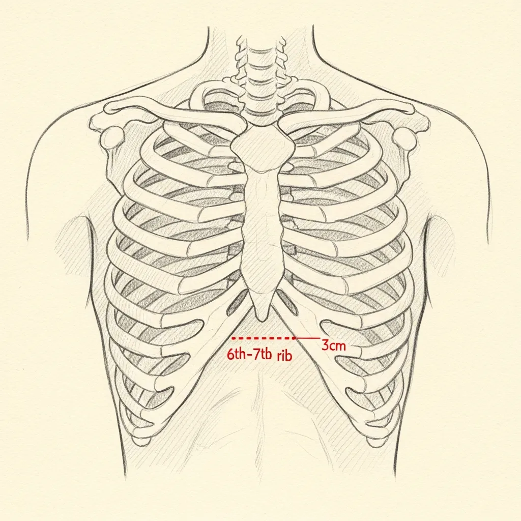
Costal
Rib cartilage for major reconstruction
Costal, or rib, cartilage is often preferred for major revision reconstruction because it can provide abundant, strong graft material when septal and auricular cartilage are depleted.
Harvest is performed through a small hidden incision in the natural inframammary crease. Donor-site discomfort, scarring, and other risks are reviewed during consultation, and pain control is individualized.
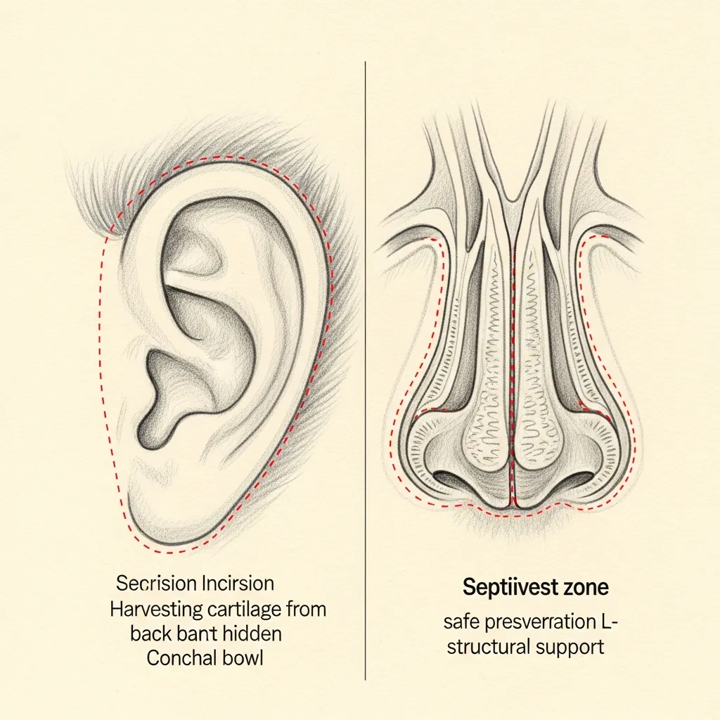
Septal & Ear
Local cartilage when supply allows
Residual septal cartilage is the first-line graft source when sufficient material remains and an L-strut can be safely preserved.
Auricular (conchal) cartilage from behind the ear provides curved, resilient graft material well-suited to lateral wall and lobular reconstruction. The harvest site is hidden in the postauricular crease.
Illustrative diagrams. Graft source is selected case-by-case based on remaining anatomy, structural need, and patient preference.
Graft Sources
Cartilage graft comparison
Revision work may require cartilage grafting or soft-tissue camouflage to rebuild what prior surgery removed or weakened. The donor source depends on how much material is needed, the load it must carry, what remains available, and the patient's preferences.
| Best use | Advantages | Limitations & risks | |
|---|---|---|---|
| Remaining septal cartilage | Small to moderate support when available | Same operative field; straight and useful for spreader grafts and smaller structural needs | Often limited after prior surgery; sufficient septal support must remain |
| Ear cartilage | Curved contour grafts, alar support, and selected tip or sidewall needs | Autologous tissue with useful natural curvature and no chest incision | Softer and naturally curved; limited quantity and not ideal for every load-bearing reconstruction |
| Autologous rib cartilage | Larger-volume or stronger reconstruction of the bridge, septum, tip, or sidewalls | Abundant autologous material that can be shaped for substantial structural needs | Requires a donor site; warping, calcification, chest-wall risks, scar, pain, and recovery must be discussed |
| Cadaveric rib cartilage | Selected cases requiring larger graft volume without a chest donor site | Avoids chest incision and harvest recovery while providing available volume | Processing, availability, surgeon preference, resorption, infection, warping, and long-term evidence require an honest discussion |
| Fascia or soft-tissue layers | Smoothing or softening visible edges in selected thin-skin or revision cases | Can camouflage an irregularity or create a softer layer over shaped cartilage | Camouflage does not replace structural support when the framework is deficient |
| Synthetic / alloplastic implant | Historically used in some nasal augmentation cases | No cartilage harvest; immediate shape support | Higher concerns for infection, extrusion, exposure, skin thinning, migration, and long-term complications in many revision contexts |
No graft source is universally best, and rib cartilage is not automatically required. Selection depends on the amount of support needed, prior surgery, donor availability, mechanical demands, tissue quality, patient preference, and the reconstructive plan.
Begin the conversation
A revision consultation is a careful, honest evaluation — not a sales conversation.
Revision Rhinoplasty Pricing Overview
Surgeon-Specific Planning Estimate
Revision Rhinoplasty With Dr. Mourad
Commonly quoted from approximately $25,000
Many cases are approximately $35,000
Estimated surgeon's fee*
Selected limited revisions may be lower after evaluation. Multiply operated, graft-intensive, implant-related, airway-reconstruction, or structurally complex cases may be higher.
* Eligible medically necessary functional components may be submitted to insurance. Cosmetic portions remain self-pay. Coverage, authorization, deductibles, coinsurance, allowed amounts, and patient responsibility vary by plan and are not guaranteed.
Final pricing follows clinical evaluation.
Revision pricing varies more widely because every prior operation changes the anatomy, scar tissue, cartilage supply, skin envelope, and degree of structural support. Factors that may increase the scope and cost include multiple prior open rhinoplasty procedures, dense scar tissue, depleted septal cartilage, nasal valve collapse, implant or graft removal, rib or ear cartilage grafting, septal perforation, major airway reconstruction, prolonged operative time, and more extensive postoperative care.
MouradNYC is an out-of-network practice. Eligible medically necessary functional components may be submitted to insurance when the patient's plan includes applicable out-of-network benefits and plan requirements are met. Cosmetic portions remain self-pay. Coverage, authorization, deductibles, coinsurance, allowed amounts, and patient responsibility vary by plan and are not guaranteed. Verification and authorization do not guarantee payment.
Published pricing is intended to provide a realistic planning estimate, not a binding quote. Every operation is individualized. Final pricing is determined only after clinical evaluation and may change if the operative plan changes.
The figures shown represent the estimated surgeon's fee. Facility, anesthesia, laboratory testing, medical clearance, imaging, prescriptions, and other services may be billed separately.
Pricing last reviewed: June 2026.
05 · In Dr. Mourad's Words
Revision rhinoplasty videos.
Short educational films and patient perspectives from the Manhattan practice.
Rhinoplasty with Dr. Mourad
An overview of Dr. Mourad's approach to rhinoplasty in his Manhattan practice.
Patient Perspective
A patient discusses her revision experience before, during, and after surgery.
Inside the Consultation
How Dr. Mourad evaluates revision anatomy, prior records, and surgical candidacy.
06 · Recovery
What revision healing actually looks like.
Stage 01
First 24 Hours
The first day centers on rest and protection of the repair. Expect congestion, support inside the nose, and some oozing managed with gentle head elevation. Pain is usually moderate and controlled with prescribed medication. Bending, lifting, and straining are avoided.
Stage 02
Week 1
The cast, when used, is typically worn for approximately one week. Swelling and bruising begin to settle after the first week, and most patients feel comfortable returning to normal social or professional activities by approximately 10–14 days.
Stage 03
Weeks 2 – 4
Exercise is gradually restarted over approximately 3–4 weeks, with higher-risk activities delayed longer. Revision tissue is scarred and less predictable, so swelling can fluctuate more than after a first-time rhinoplasty.
Stage 04
Months 1 – 18
Meaningful improvement occurs over the first several months, but final refinement commonly takes 12–18 months. Particularly complex or thick-skinned revision cases may continue evolving for 18–24 months.
Have a specific question?
Send a brief note describing your anatomy or concerns — the office will route it directly to Dr. Mourad for review.
Twelve to Eighteen Months
Long-term healing.
Outcomes after revision rhinoplasty evolve across predictable phases that reflect healing, graft integration, and scar remodeling rather than a single endpoint. The early phase is characterized by edema, bruising, and firm tissues that obscure fine contours. The intermediate phase entails graft settling, soft-tissue relaxation, and improving nasal symmetry as inflammation resolves. The late phase, typically approaching twelve months, reveals more stable tip definition and dorsal refinement, though subtle changes may continue for longer in complex reconstructions. Because revision cases usually involve added structural work, the surgeon anticipates gradual maturation and plans follow-up accordingly.
Functional improvements in airway patency follow a similar multi-stage course and depend on both mechanical repair and mucosal recovery. Septal reconstruction, turbinate reduction, and valve support procedures reduce structural blocks to airflow, but mucosal swelling and scar contracture influence subjective breathing for months. Objective assessment with nasal endoscopy, validated patient-reported outcome measures, or airflow testing at follow-up visits helps track progress in both subjective and measurable terms. Successful functional outcomes therefore rely on combined surgical mechanics and postoperative medical care tailored to mucosal health.

Safety & Consent
Risks, safety, and informed consent.
Revision rhinoplasty carries specific risks that differ from primary procedures because scarred tissue and prior grafting change the operative landscape. Bleeding and hematoma formation remain early risks and can compromise grafts or require urgent drainage in the operating room. Septal hematoma (a collection between nasal septum layers) produces worsening pain and obstruction and requires prompt drainage to avoid cartilage loss. Mitigation includes meticulous intraoperative hemostasis, perioperative blood-pressure control, and early postoperative monitoring for expanding nasal fullness or new drainage that suggests a hematoma.
Infection is a recognized complication that may present with fever, increasing pain, erythema, and purulent drainage from incision or donor sites. Alloplastic implants have a higher risk of late exposure and infection compared with autologous grafts, and implant-related infections sometimes mandate removal. To reduce infectious risk we apply strict sterile technique, judicious antibiotic use when indicated, and selection of autologous tissue when feasible. Early identification of infection allows office-based irrigation and targeted antibiotics or timely operative washout when necessary to protect structural grafts.

Before You Arrive
Your revision consultation, prepared.
Bring prior operative reports, including records of septal cartilage harvest or airway work, when available.
Bring preoperative and postoperative photographs from each prior nasal procedure when available.
Bring implant or graft information, pathology reports, and relevant imaging. These may clarify what was removed or placed and which structures may still be usable.
Missing records do not prevent evaluation, but they can increase uncertainty; examination, photography, endoscopy, or selective imaging may still help define the current anatomy.
Note any breathing changes since your prior surgery — when, how severe.
List all medications, supplements, and blood-thinning agents.
Allow 75 minutes; revision consultations are deliberately longer.
No decisions are made at the first visit — that is by design.
In Their Words
From patients of the practice.
Revision surgery made me nervous because I had already been through one rhinoplasty elsewhere. This time felt very different. Everything was explained clearly, and the result looks much more balanced.
I had breathing issues and cosmetic concerns after my first surgery years ago. My revision was not rushed, and I appreciated the honesty about what could and couldn't be fixed. My nose looks softer and functions better.
I wanted a revision but was scared of making things worse. The plan was conservative, and that gave me confidence. I finally feel like my nose fits my face.
I flew in for revision rhinoplasty after several consultations that didn't feel right. This was the first time I felt someone really understood the problem. The improvement has been a huge relief.
Individual experiences. Results and recovery vary by patient. Testimonials shared with written consent.
Patient Reviews
Revision Rhinoplasty Patient Experiences
Selected public patient reviews. Individual experiences vary.
“After a really difficult first surgery, I was hesitant about revision rhinoplasty, until I met Dr. Mourad. He was incredibly warm and attentive from the start…”
Real patient experiences
Selected public reviews from patients of the practice.
Your privacy matters
We never share personal health information.
Board-certified expertise
Dual board-certified facial plastic and reconstructive surgeon.
Individual results vary. Reviews reflect individual experiences and are not a guarantee of outcome.
At a Glance
Revision rhinoplasty fact snapshot.
- Treats
- Structural collapse, breathing obstruction, asymmetry, over-resection, tip deformity, and bridge irregularities after a prior rhinoplasty.
- Does not treat
- Concerns unrelated to the prior surgery, or expectations of an entirely different nose — revision works within the limits of the remaining tissue.
- Evaluation
- In-person examination of the internal and external nose, review of prior operative records when available, and sometimes imaging.
- Related conditions
- Nasal valve collapse, deviated septum, and post-surgical airway obstruction.
- Possible combined procedures
- Septoplasty, nasal valve repair, and cartilage grafting from the septum, ear, or rib.
- Recovery summary
- Most patients return to desk work in one to two weeks; swelling resolves gradually over a year or more, often slower than after a primary rhinoplasty.
- Insurance / functional distinction
- The functional airway component may be submitted for insurance review — coverage is never guaranteed; cosmetic revision is self-pay. This out-of-network practice provides documentation for reimbursement.
- When to seek evaluation
- Once healing has stabilized — typically around a year after the prior surgery — or sooner for significant breathing obstruction.
Find your starting point
Which nasal breathing path fits your situation?
Answer three short questions to see which page explains the evaluation most relevant to you. This is educational orientation, not a diagnosis — a clinical examination is how the cause of nasal obstruction is actually determined.
Select an option for each question to see which page fits your situation.
This guide is general educational information, not medical advice or a diagnosis. Only an in-person examination can determine the cause of nasal obstruction and the appropriate treatment.
See all pathways
A combined revision evaluation likely fits
When both breathing and appearance are concerns after previous surgery, the anatomy is already altered and rebuilding structural support is often part of the plan. A revision evaluation looks at what the prior operation changed before anything is recommended.
A revision rhinoplasty conversation likely fits
Changing the appearance of a nose that has already had surgery is a different undertaking from a first operation, because scar tissue and prior changes shape what is possible. A revision evaluation is the right starting point.
A deviated-septum evaluation likely fits
Persistent, constant blockage that does not resolve with sprays or allergy treatment is often related to a deviated septum. Septoplasty is the functional operation that straightens the septum. If you have had prior nasal surgery, revision considerations also apply.
A nasal valve evaluation likely fits
When the side of the nose draws inward on hard inspiration, the nasal valve — the narrowest part of the airway — may be the contributor rather than the septum alone. This is assessed directly on examination.
A turbinate and sinus evaluation likely fits
Congestion that fluctuates with colds, allergies, or facial pressure often involves swollen turbinates or the sinuses rather than the septum by itself. The evaluation looks at the whole airway before any procedure is considered.
A septorhinoplasty conversation likely fits
When breathing and appearance are both concerns, they can often be addressed together in a single planned operation — straightening the septum while refining the external shape — so there is one recovery rather than two.
A rhinoplasty conversation likely fits
When the concern is the shape of the nose, rhinoplasty is the operation that refines it. A consultation focuses on your goals and whether the underlying anatomy supports them.
A revision evaluation likely fits
After previous nasal surgery, a fresh structural evaluation is the right starting point because the anatomy has already been changed once.
Start with a functional nasal evaluation
Your answers point to more than one possible contributor, which is common. A functional nasal examination is how the cause is identified before any treatment is considered.
Why patients trust this practice
Care led by a double board-certified specialist
Double board certified
American Board of Facial Plastic & Reconstructive Surgery and American Board of Otolaryngology — Head & Neck Surgery.
AAFPRS Fellowship Director
Trains fellows through the American Academy of Facial Plastic and Reconstructive Surgery.
Published author
Contributions to the academic literature of facial plastic surgery.
Face, nose & sinus focus
A practice concentrated above the clavicles, including complex revision evaluations.
Frequently Asked
Revision Rhinoplasty NYC FAQs
Revision rhinoplasty cost in NYC is highly individualized because the surgeon may be working with scar tissue, altered anatomy, depleted cartilage, breathing obstruction, or the need for grafting. There is no flat price; the fee reflects the surgical complexity, anesthesia, and facility costs for your specific case. A personalized estimate is provided after an in‑person consultation.
Cosmetic refinements are generally self‑pay. If part of the revision corrects a documented functional problem such as nasal obstruction, valve collapse, or septal deviation, insurance may apply to the functional portion. Coverage depends on your plan, medical necessity, and documentation. Our office can help you understand what records may be needed and what out‑of‑pocket costs may apply.
Clinical references
This page draws on published clinical practice guidelines and public-health references. These sources inform general patient education and do not replace an individual evaluation with Dr. Mourad.
- 01Ishii LE, et al. Clinical Practice Guideline: Improving Nasal Form and Function after Rhinoplasty. Otolaryngology–Head and Neck Surgery. 2017;156(2 Suppl):S1–S30. PMID: 28145823. DOI: 10.1177/0194599816683153. AAO-HNSF guideline
- 02Rohrich RJ, et al. Fresh Frozen Rib Cartilage Grafts in Revision Rhinoplasty: A 9-Year Experience. Plastic and Reconstructive Surgery. 2022;150(1):58–62. PMID: 35511072. DOI: 10.1097/PRS.0000000000009203. PubMed
- 03Datta S, et al. Rib Grafts in Rhinoplasty: A Systematic Review and Meta-analysis of Autologous, Fresh-frozen, and Irradiated Cartilage. Plastic and Reconstructive Surgery Global Open. 2026;14(6):e7875. PMID: 42376269. DOI: 10.1097/GOX.0000000000007875. PubMed
- 04Robotti E, et al. The Evolution of the SPF-SPLF Graft in Rebuilding the Dorsum in Revision Rhinoplasty—an Update in an Over 300 Case Series. Aesthetic Plastic Surgery. 2026;50(1):130–139. PMID: 40826294. DOI: 10.1007/s00266-025-05160-9. PubMed
- 05Chen K, Zhou L. The Effect of Functional Rhinoplasty on Quality of Life: A Systematic Review and Meta-Analysis. Aesthetic Plastic Surgery. 2024;48(5):847–854. PMID: 37173413. DOI: 10.1007/s00266-023-03390-3. PubMed
Related Nasal Surgery
Explore related procedures and resources.
Revision is the right path only for patients with prior nasal surgery. If your situation is different, these pages may be a better starting point.
Primary rhinoplasty in NYC
For patients who have never had nasal surgery and are considering a first rhinoplasty.
Read moreSeptoplasty for a deviated septum
When the primary concern is breathing through a deviated septum rather than appearance.
Read moreNasal valve collapse treatment
Repair of internal or external valve collapse that restricts the nasal airway.
Read moreSeptal perforation repair
Closure of a hole in the septum, sometimes sustained during prior nasal surgery.
Read moreBroken nose repair
Evaluation and repair after recent nasal trauma or a post-traumatic deformity.
Read moreRevision rhinoplasty cost in NYC
What affects revision surgery fees — surgeon, facility, anesthesia, rib or ear cartilage grafting, and insurance.
Read moreCost & insurance
How surgical fees and insurance for functional repair are approached.
Read moreThe Most Important Step
Your revision consultation.
The appropriate next step is an in-person consultation where prior operative reports, photographs, and imaging are reviewed to shape an individualized plan. Bring all prior surgical records, implant documentation if available, and a clear chronology of functional and cosmetic symptoms to the visit. Expect the consultation to include an external exam and office nasal endoscopy (a flexible scope examination) to assess mucosal health, septal integrity, and valve competence. We will discuss candidacy, whether septal cartilage remains available, the potential need for auricular or costal cartilage, and whether a staged reconstruction is safer for durable results.